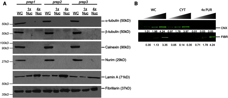
Fig. 6.
Immunoblot analyses of nuclear fractions. A: ECL immunoblot analysis of whole cell (WC) and purified nuclear fractions (NUC). Fractions collected from three separate nuclear preparations were prepared for gel electrophoresis as described in the Materials and Methods. Gels were loaded by cell equivalents based on the amount of whole cell lysate needed for blotting in the linear range. Comparison to a 4-fold excess of purified nuclei (4× PUR) highlights the absence/enrichment of cellular proteins in nuclear fractions. B: Odyssey immunoblot analysis of calnexin and fibrillarin content in whole cell, cytosolic, and 4× nuclear fractions from a single preparation. Each sample was loaded in triplicate. Gels were loaded with 15,000, 30,000, and 60,000 cell equivalents of both whole cell (WC) and cytosolic (CYT) protein fractions, whereas 60,000, 120,000, and 240,000 cell equivalents of purified nuclear protein were loaded for analysis. Relative intensity of each individual band was quantified using the Odyssey 2.0 software. These values are listed below the image underneath the corresponding bands. CNX, calnexin; FIBR, fibrillarin.

