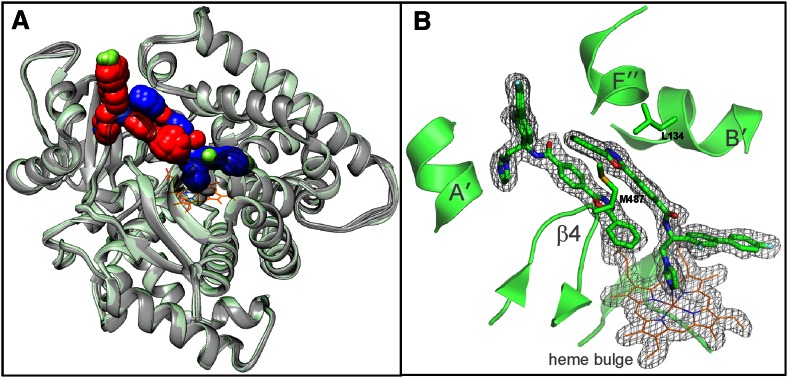
Fig. 4.
Crystal structures of human CYP51 in complex with VFV. A: Superimposition of four monomers from 4UHI and eight monomers from 4UHL; rms deviation for the Cα atom positions 0.34 ± 0.06 Å. One heme molecule (orange lines) is outlined for clarity. All VFV molecules are shown as sphere representation. The C-atoms in the heme-coordinating VFV molecules are blue and in the substrate entry shielding VFV molecules are red. B: The 2Fo-Fc omit electron density map around the CYP51 heme and two VFV molecules, contoured at 1.5 σ. Orientation is as in part (A). Some secondary structural elements and the pair of substrate channel gating residues (Met484 and Leu134) are also shown.

