Fig. 2.
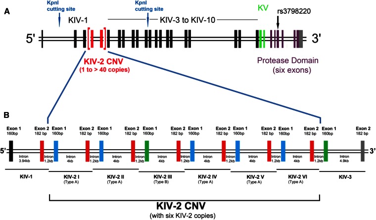
A: Exon-intron structure of different domains of the human LPA gene. Each KIV domain (KIV-2 in red, KIV-1 and KIV-3 to KIV-10 shown in black) and KV (green) consists of two exons, while the PD (purple) has six exons. The location of the KpnI cutting sites and the nonsynonymous SNP, rs3798220, is shown (137, 157). Modified from (201). B: Exon-intron structure of the KIV-2 domain and directly flanking KIV-1 and KIV-3 domains. The KIV-2 CNV is shown according to the LPA reference sequence (ENSG00000198670; GRCh38), which contains six KIV-2 copies. Each KIV-2 copy has a size of 5.5 kb and consists of two exons separated by a long intron of 4 kb. A short intron of 1.2 kb separates the two KIV-2 copies. Exon 2 (182 bp) of each KIV-2 copy is identical (red). Exon 1 (160 bp) is of three different types: type A (shown in blue), type B (shown in green), and type C (not depicted here). Types A, B, and C of exon 1 are differentiated by synonymous mutations. The third KIV-2 copy in the LPA reference sequence (ENSG00000198670; GRCh38) contains exon 1 of type B and is shown here accordingly. Note that the type B exon 1 of KIV-2 is 100% identical in its nucleotide sequence to KIV-3 exon 1 (both shown in green), and KIV-2 exon 2 has 100% identical nucleotide sequence as KIV-1 exon 2 (shown in red). This figure is not drawn to the scale.

