Figure 1.

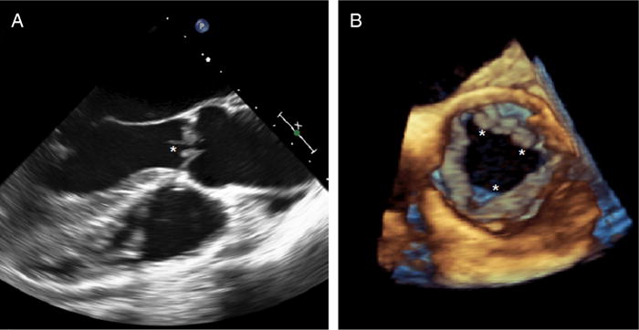
Transoesophageal echocardiogram (TEE) revealing multiple densities (*) attached to the ventricular surface of the aortic valve, seen in 2D TEE (long-axis view, A) and 3D TEE (short-axis view, B).

Transoesophageal echocardiogram (TEE) revealing multiple densities (*) attached to the ventricular surface of the aortic valve, seen in 2D TEE (long-axis view, A) and 3D TEE (short-axis view, B).