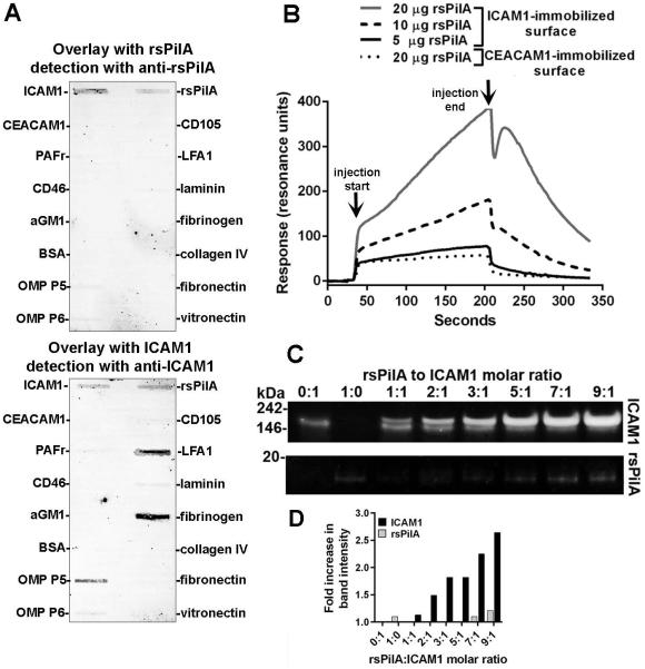
Figure 1.
rsPilA bound to ICAM1. (A) Far Western slot blot wherein incubation of rsPilA (top blot) or ICAM1 (bottom blot) with a panel of host cell receptors and extracellular matrix proteins adsorbed on to PVDF membranes revealed reactivity of rsPilA exclusively to ICAM1. Conversely, ICAM1 interacted with rsPilA, in addition to its known binding partners LFA1, NTHI OMP P5 and fibrinogen. (B) Sensorgram curves for surface plasmon resonance wherein ICAM1 immobilized to the surface of a sensor chip was exposed to rsPilA demonstrated a concentration-dependent increase in reactivity, a response not observed when rsPilA (at the greatest concentration) was assayed versus a CEACAM1-immobilized control surface. Arrows indicate start and stop of the injection cycle. (C) An increase in ICAM1 band intensity was shown by native PAGE upon incubation of ICAM1 with increasing molar ratios of rsPilA, a result quantitated in (D). Collectively, these data demonstrated that rsPilA bound to ICAM1.

