Figure 4.
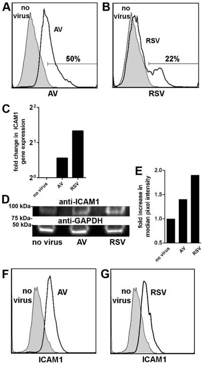
Inoculation of polarized epithelial cells with RSV or adenovirus resulted in increased expression of ICAM1. Incubation of NHBEs with (A) adenovirus or (B) RSV at MOI 2 for 72 h yielded positive labeling for respective viral antigen (clear histograms), compared to untreated cells (shaded histograms) as determined by flow cytometry. The proportion of cells that expressed viral antigen is indicated. (C) Fold change in ICAM1 gene expression normalized to GAPDH gene expression and relative to uninfected cells revealed a relative increase in ICAM1 transcript abundance by virus-infected NHBEs. (D) SDS-PAGE and western blot analysis of cell lysates probed for ICAM1 demonstrated an increase in band intensity after viral infection compared to uninfected cultures and (E) was quantitated by densitometry. Equivalent protein concentration per sample was indicated by probing for GAPDH (D, bottom row). ICAM1 expression increased on NHBEs infected with adenovirus (F, clear histogram) or RSV (G, clear histogram) compared to uninfected cells (F & G, shaded histograms).

