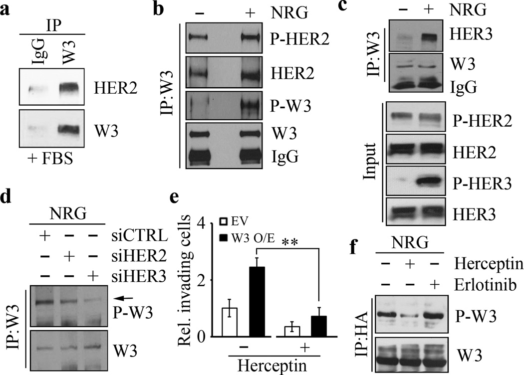
Figure 3. WASF3 is phosphorylated following NRG stimulation through binding to the HER2/HER3 heterodimer.
IP of WASF3 in the presence of serum leads to co-precipitation of HER2 (a) in SKBR3 cells. In the presence of NRG, there is only a modest increased in activated HER2 levels but the majority of the WASF3 protein in the immunocomplex is phospho-activated (b). IP of WASF3 in SKBR3 cells shows the presence of high levels of HER3 in the immunocomplex (c). Analysis of the NRG-treated SKBR3 cells shows only a modest increase in HER2 levels but a dramatic increase in activated HER3 (c). IP of WASF3 shows a decrease in WASF3 phosphorylation levels (d) either in HER2 knockdown (siHER2) or HER3 knockdown (siHER3) SKBR3 cells compared with the knockdown control cells (siCTRL). Treatment of SKBR3 cells overexpressing WASF3 with Herceptin shows a significant reduction in the invasion potential (e). Herceptin treatment of WASF3-overexpressing SKBR3 cells leads to a reduction in levels of activated WASF3, which is not seen in cells treated with the Erlotinib suppresser of EGFR signaling (f). ** p<0.01; Student’s t-test.

