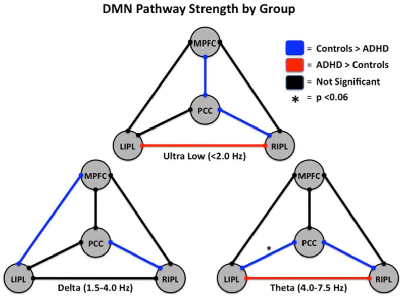
Figure 4. Functional Connectivity in the Default-Mode Network of Adults with and without ADHD.

Three models of the default-mode network (DMN) showing group differences and similarities in functional connectivity along particular pathways at distinct frequencies of neuronal activity are shown. See Figure Legend (upper right) for definition of pathway colors. In the ultra-low frequency range, adults without ADHD showed stronger coupling between the medial prefrontal cortices (MPFC) and posterior cingulate cortices (PCC) and the PCC and right inferior parietal (RIPL) areas, whereas those with ADHD showed stronger connectivity between the RIPL and left inferior parietal (LIPL) nodes. In the delta band, controls showed increased connectivity between the MPFC and LIPL regions and the PCC and RIPL regions relative to un-medicated patients with ADHD. Theta frequency neuronal activity was more strongly coupled between the PCC and the RIPL and LIPL areas in adults without ADHD relative to their un-medicated peers with ADHD, while the latter group showed stronger coupling between the RIPL and LIPL cortical areas. Interestingly, the increased functional connectivity in the PCC-RIPL pathway of adults without ADHD was consistent throughout all three frequency ranges of neuronal coupling. Following medication administration, all differences in connectivity between adults with and without ADHD dissipated and were no longer significant, indicating that stimulants increased connectivity along some pathways and decreased it along others.52
