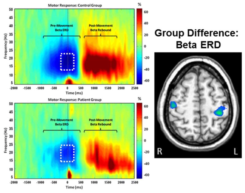
Figure 7. Aberrant Beta Oscillations in Parkinson's Disease (PD).

(Left) Average time-frequency spectrograms from a MEG sensor near the sensorimotor cortex are shown for healthy controls (top panel) and patients with PD (bottom panel). Time (in ms) is denoted on the x-axis, with 0 ms defined as movement onset. Frequency (in Hz) is shown on the y-axis. The typical pattern of peri-movement beta desynchronization (ERD) and post-movement beta rebound (PMBR) during a right-hand movement task, expressed as percent difference from baseline, can be discerned in each group. The time period within the white box (-300 to 200 ms) was subjected to source reconstruction analyses. These analyses revealed that peak group differences in the beta ERD response (right) were within the motor hand knob region of the left precentral gyrus and extended onto the adjacent postcentral gyrus (p = .035, corrected). In both cases, responses were weaker in patients with PD. Trending differences were also seen in the right precentral gyrus near the hand knob region (p < .005, uncorrected; p = .107, corrected).113 Abnormalities in beta activity appear to be a central feature of PD and have been observed in the cortex in MEG studies and in the subthalamic nucleus during deep brain stimulation (DBS) surgeries.
