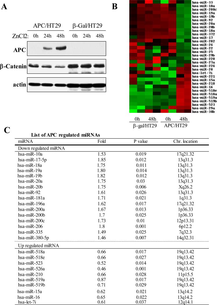
Figure 1. Profile of APC-regulated miRNAs.
(A) Western blot. APC/HT29 and β-Gal/HT29 cells were treated with ZnCl2 for indicated times and then subjected to immunoblot analysis with indicated antibodies (note: expression of APC leads to decrease in β-catenin level). (B and C) Heatmap (B) and table (C) show the miRNAs significantly regulated by expression of APC.

