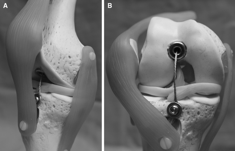
Fig. 6.
Schematic model of the CDS implanted into the right femur. a Antero-medial view at 10° flexion of the knee joint. The traction wire is fixed to the tibial tubercle proximal to the insertion of the patellar ligament. b Anterior view at 90° flexion of the knee joint. The patella is laterally dislocated to fully expose the intra-articular running of the wire. The traction wire and the distal end of the nail do not impinge the menisci or impact on the ACL and the retro-patellar cartilage

