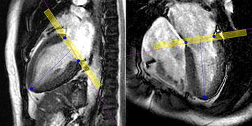
Fig. 2.

Use of the long-axis views for LV volumetry from SSFP imaging. The operator manually specified the basal end and apex of the left ventricle (blue dots) on the vertical (left) and horizontal (right) long-axis views. The basal slice contained the left atrium and LV even within a pixel, and the processing using the long-axis images aided extraction of the true LV volume. The yellow box, representing the volume of the basal slice, was added for explanation to the actual display of the cvi42 software
