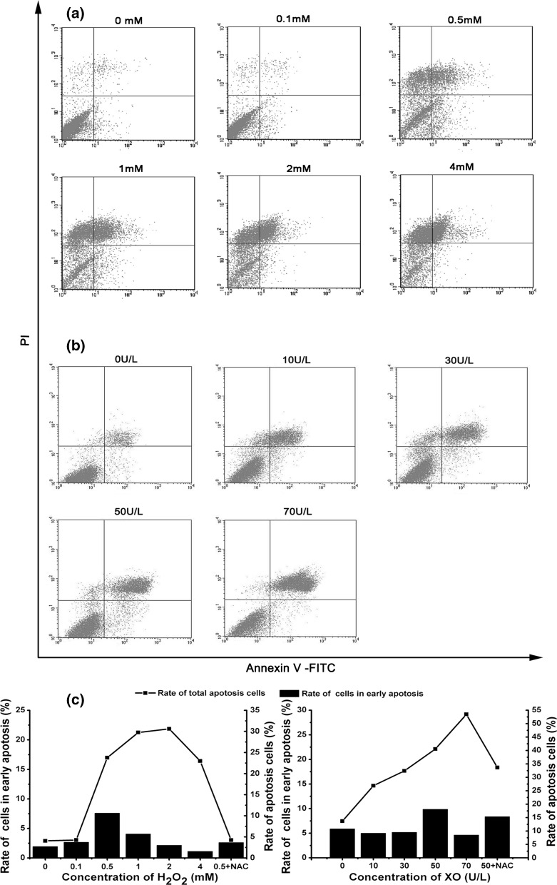
Fig. 3.
Apoptosis of IPEC-J2 cells as determined by flow cytometry after coincubation with H2O2 or with X/XO. The apoptotic rates were analyzed by Annexin V/PI staining. a Apoptosis of IPEC-J2 cells incubated with different concentrations of H2O2; b apoptosis of IPEC-J2 cells incubated with 250 μM X and with different concentrations of XO for 6 h; c rate of early apoptotic IPEC-J2 cells (annexin+, PI−; quadrants 4) or total apoptotic cells (annexin+; quadrants 1, 4)

