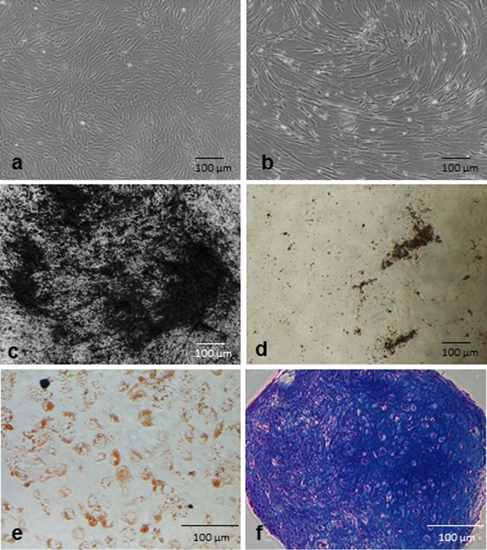
Fig. 5.
Representative photographs of cell morphology and differentiation. Morphology of passage 2 cells was normal spindle shaped for both a PL1- and b PL2-medium cultured BM-MSCs, but the PL2-cultured cells appeared to have a slightly larger morphology. Von Kossa staining of osteogenic differentiated BM-MSCs in c PL1-supplemented differentiation medium and in d FBS-supplemented differentiation medium. e Sudan III staining indicated the adipogenic differentiation potential of the samples and f Alcian blue staining indicated the chondrogenic potential of the cells grown in PL1-medium

