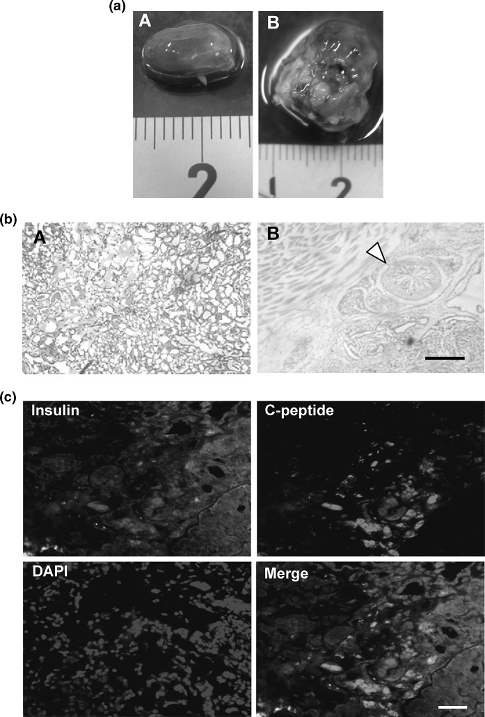
Fig. 6.
Histochemical analytical results. a Effect of the removal of SSEA-1 positive cells on the formation of teratoma. (A) A kidney taken out at the star in Fig. 5a. (B) A kidney taken out from another mouse in which the produced cells were transplanted separately without removal of SSEA-1 positive cells. b Cell morphology revealed by HE staining. (A) a section prepared from (aA). (B) a section prepared from (aB). A triangle indicates an intestine-like structure. Scale bar indicates 500 μm. c Fluorescent images of insulin, C-peptide, and nucleus of a section prepared from (aA). Scale bar indicates 50 μm. (Color figure online)

