Fig. 1.
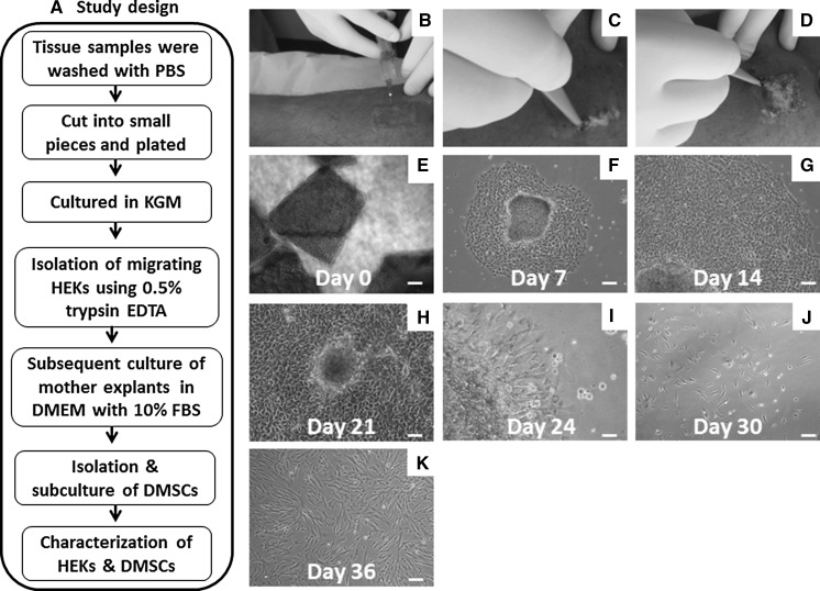
Isolation and propagation of HEKs and DMSCs from human skin biopsies: a Flow chart describes study design. b–d Surgical procedures for obtaining skin biopsy samples. Phase contrast photographs showing; e initial plating of minced skin samples with growth medium. f–h Outgrowth of HEKs showing polygonal morphology. i secondary outgrowth of stromal phenotypes from mother explant which were left intact after trypsinization. j–k Scattered growth of stromal cells with spindle morphology. Scale bars 100 μm (e–k). (Color figure online)

