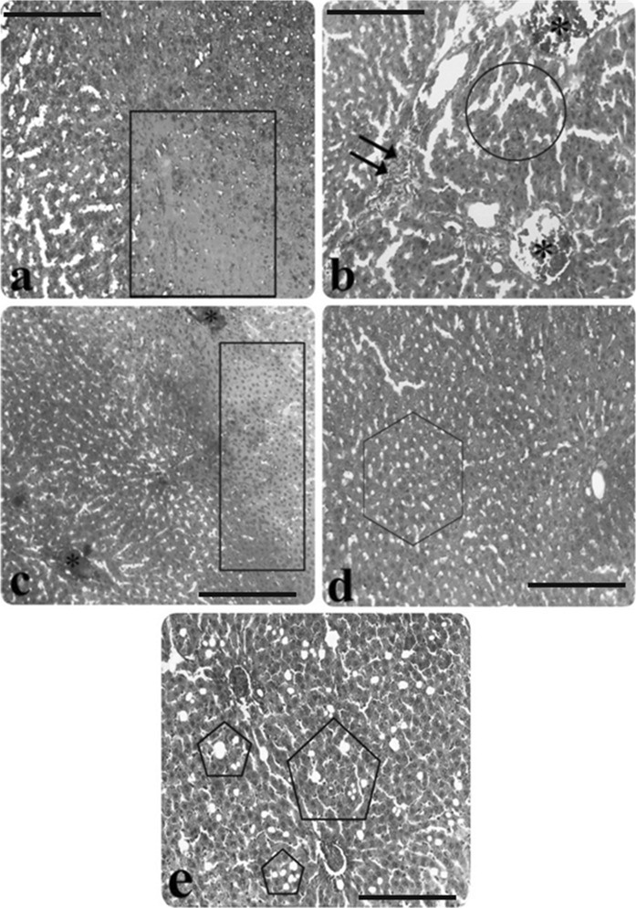
Fig. 2.
Light microscopic appearance of liver from AP group rats. a Hepatocyte necrosis (inside of the square symbol), b) Infiltration (double arrow), congestion (asterisk), sinusoidal dilatation (inside of the circle symbol), c hepatocyte necrosis (inside of the rectangular symbol), congestion (asterisk), d vacuolisation (inside of the hexagon symbol), e edema (inside of the pentagon symbol), (H&E staining). Scale bars in a, b, c, d, e 50 µm

