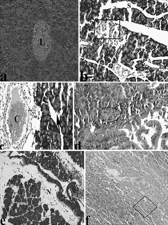
Fig. 1.
Light microscopic appearances of pancreas from control and AP rats. a Pancreas of control group, Langerhans islet (L). Pancreas in AP rats; b major Sinusoidal dilatation (S), degeneration of Langerhans (inside of quadrangle symbol), c congestion (C), asinus necrosis (black arrow), d infiltration (I), fibrosis (inside of circle symbol), portal vein (Pv), e fat necrosis (N), edema (E), f vacuolisation (inside of square symbol), (H&E)

