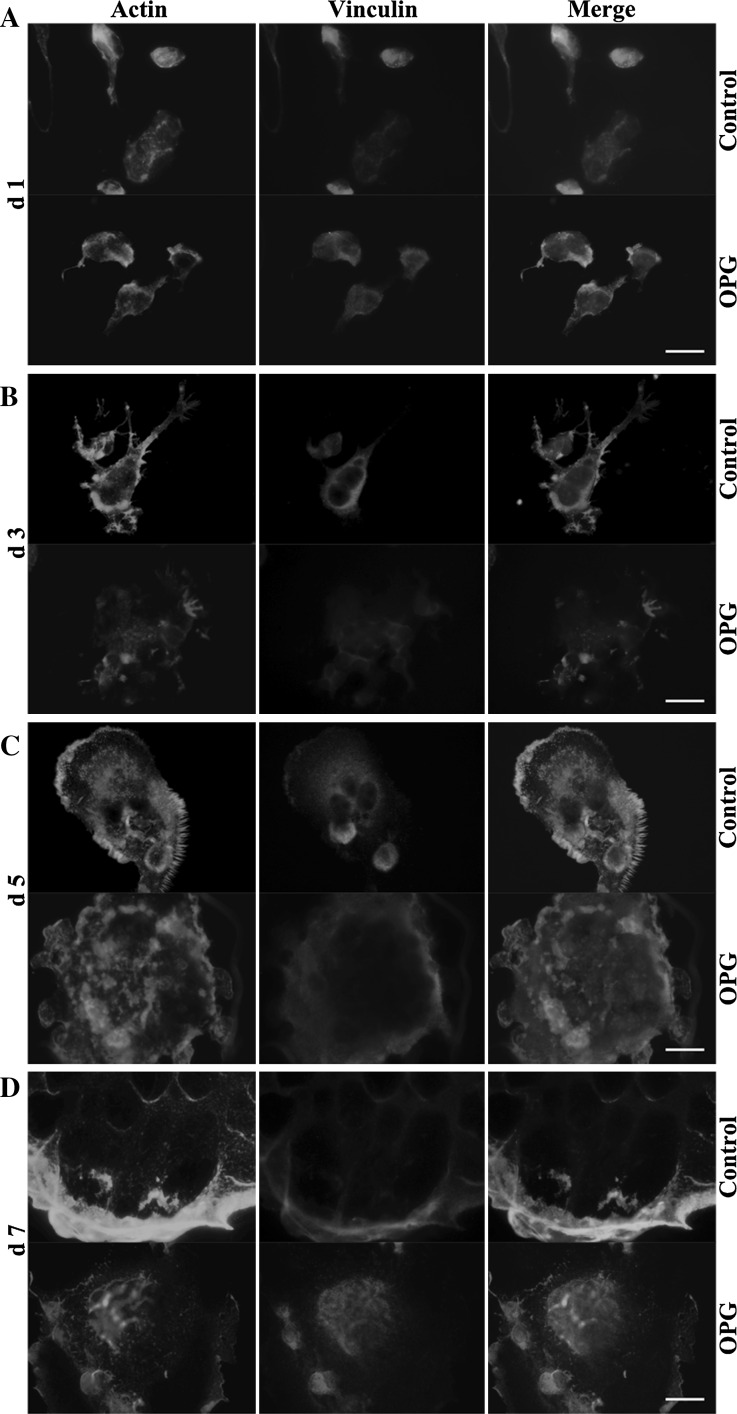
Fig. 2.
OPG exposure at different time points differentially regulates the formation of podosomes and adhesion structures. RAW264.7 cells were seeded on coverslips and cultured for 1 (A), 3 (B), 5 (C), or 7 (D) days, as described in the “Methods” section. Cells were treated with 80 ng/mL OPG for 24 h (lower panels) or left untreated (upper panels) and then were fixed and stained to detect F-actin (red), vinculin (green), and nuclei (blue), and samples were imaged using a fluorescent microscope (magnification ×1000, scale bar = 10 μm). (Color figure online)

