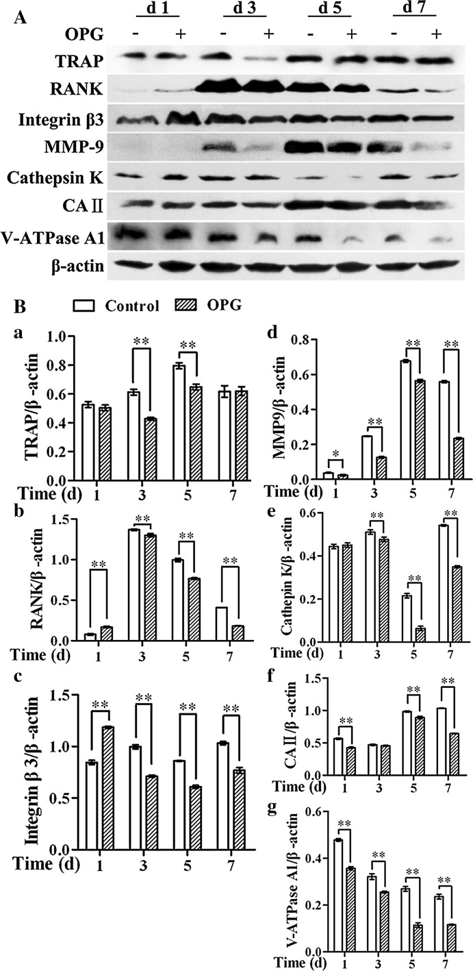
Fig. 3.
OPG exposure at different points differentially regulates the expression of osteoclast enzymes and markers. A Cell lysates were immunoblotted with the indicated antibodies and analyzed by western blotting. B The band intensities were quantified by densitometry, and β-actin was used as a loading control. The results are expressed as the mean ± SD. **p < 0.01 and *p < 0.05 versus control

