. 2016 Jul 1;12(3):262–273. doi: 10.3988/jcn.2016.12.3.262

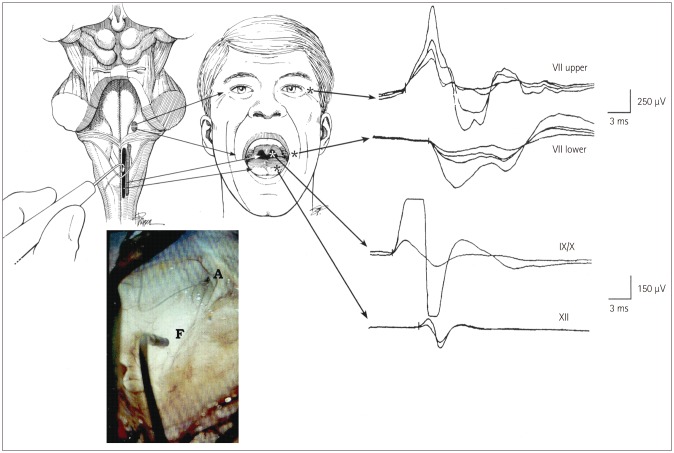
Fig. 1. Mapping of the brainstem cranial nerve motor nuclei (CMN). Upper left: drawing of the exposed floor of the fourth ventricle with the surgeon's hand-held stimulating probe in view. Upper middle: sites where hook-wire electrodes were inserted for recording muscle responses. Far upper right: compound muscle action potentials recorded from the orbicularis oculi and oris muscles after stimulation of the upper and lower facial nuclei (upper two traces), and from the pharyngeal wall and tongue muscles after stimulation of the CMN of cranial nerves IX, X, and XII (lower two traces). Lower left: photograph obtained with the aid of the operating microscope showing the hand-held stimulating probe placed on the floor of the fourth ventricle (F). A: aqueduct. Adapted from Morota et al., Neurosurgery 1995;37:922-929; discussion 929-930, with permission from Wolters Kluwer Health, Inc.10.

Fig. 1