. 2016 Jul 1;12(3):262–273. doi: 10.3988/jcn.2016.12.3.262

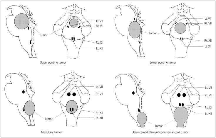
Fig. 2. Typical patterns of CMN displacement by brainstem tumors in different locations. Upper and lower pontine tumors typically push the facial nuclei around the edge of the tumor, suggesting that it is necessary to precisely locate the facial nuclei before tumor resection in order to avoid them being damaged during surgery. Medullary tumors typically grow more exophytically and compress the lower CMN ventrally; these nuclei may be located on the ventral edge of the tumor cavity. Because of the interposed tumor, in these cases mapping before tumor resection usually does not allow identification of the CMN of cranial nerves IX, X, and XII. However, responses may be obtained close to the end of the tumor resection after most of the tumor tissue between the stimulating probe and the motor nuclei has been removed. Repeated mapping is recommended at this point since the risk of damaging motor nuclei is significantly higher than at the beginning of tumor debulking. Cervicomedullary junction spinal cord tumors simply push the lower CMN rostrally when they extend into the fourth ventricle. Adapted from Morota N, et al., Neurosurgery 1996;39:787-793; discussion 793-794, with permission from Wolters Kluwer Health, Inc.8 CMN: cranial nerve motor nuclei.

Fig. 2