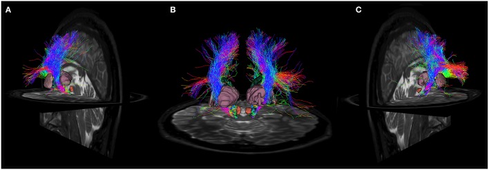
Figure 5.
Anatomical relations of cortico-nigral probabilistic fiber tracking. In particular, panel (A) depicts left cortico-nigral pathway in a posterior parasagittal view; panel (B) shows bilateral cortico-nigral tract in posterior coronal view and panel (C) shows right cortico-nigral tract in posterior parasagittal view. 3D-rendered models of basal ganglia are shown to better illustrate anatomical relation: SN is shown colored in blue, caudate nucleus in dark green, thalamus in pale purple, putamen in pink, pallidum in light blue, STN in yellow and RN in brown. Cortico-nigral tracts runs mainly through internal capsule, avoiding thalamus, putamen and globus pallidus, and reach SN in the ventral midbrain, bypassing STN and RN.

