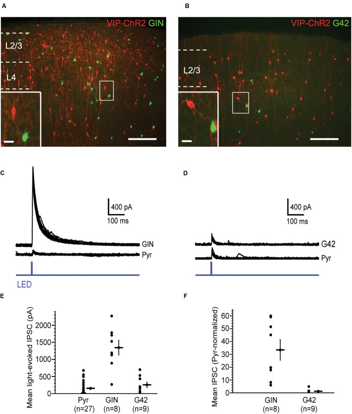
FIGURE 3.
Vasoactive intestinal peptide (VIP) cells strongly inhibit SOM cells. (A) Fluorescent image of barrel cortex of GIN mouse (endogenous GFP in subset of SOM cells) crossed with VIP-Cre mouse, virally transfected with Cre-dependent, RFP-tagged ChR2; scale bar = 200 μm. Inset: expanded view of neighboring VIP cell (red) and GIN cell (green); scale bar = 20 μm. (B) Fluorescent image of barrel cortex of G42 mouse (endogenous GFP in subset of PV cells) crossed with VIP-Cre mouse, virally transfected with Cre-dependent, RFP-tagged ChR2; scale bar = 200 μm; dotted line denotes pia. Inset: expanded view of neighboring VIP cell (red) and G42 cell (green); scale bar = 20 μm. (C) Light-evoked IPSCs in a GIN cell and neighboring pyramidal cell in layer 2/3, in which VIP cells express ChR2. 10 traces overlaid for both GIN and pyramidal cell. (D) Light-evoked IPSCs in a G42 cell and neighboring pyramidal cell in layer 2/3, in which VIP cells express ChR2. 10 traces overlaid for both G42 and pyramidal cell. (E) Population data of mean light-evoked IPSCs in pyramidal, GIN, and G42 cells when VIP cells are optogenetically activated. IPSCGIN > IPSCPyr (P = 1.1 × 10⁻⁵, Kruskal–Wallis test, Bonferroni correction), IPSCGIN > IPSCG42 (P = 4.3 × 10⁻⁴, Kruskal–Wallis test, Bonferroni correction). (F) Population data of mean light-evoked IPSCs in GIN, and G42 cells, normalized to mean light-evoked IPSCs in pyramidal cells in same slice, when VIP cells are optogenetically activated, normIPSCGIN > normIPSCG42 (P = 4.4 × 10⁻⁴, Mann–Whitney test).

