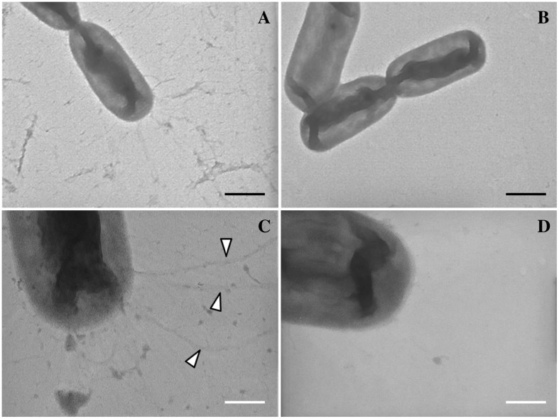
FIGURE 6.
Visualization of type IV pili of Lysobacter capsici AZ78 using Transmission Electron Microscopy (TEM). Pilus-like structures (arrows) emerge at the pole of L. capsici AZ78 cells grown on Pea Agar Medium amended with 0.5% Agar (w/v; A,C) and these structures are absent in cells grown on Luria Bertani Agar amended with 0.5% Agar (w/v; B,D). A and B, magnification X-19000, black bar 500 nm. (C,D), magnification X-64000, white bar 150 nm.

