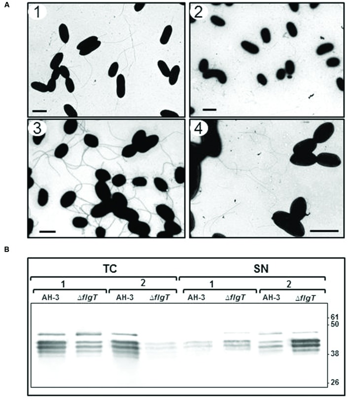
FIGURE 4.
(A) Transmission electron microscopy of AH-3ΔflgT mutant during the mid-log-phase (OD600 ≈ 0.5) (1) and the late-log-phase (OD600 ≈ 2) (2) growth at 25°C on liquid media and at the late-log-phase growth in soft agar plates (3). A. hydrophila AH-3 during the late-log-phase growth at 25°C on liquid media (4). Bacteria were gently placed onto Formvar-coated copper grids and negatively stained using 2% uranyl acetate. Bar = 2 μm. (B) Western-blot of total bacterial cells (TC) and supernatants (SN) of A. hydrophila AH-3 and AH-3ΔflgT mutant during the mid-log-phase (1) and the late-log-phase (2) growth at 25°C on liquid media, using specific antiserum against purified polar flagellins.

