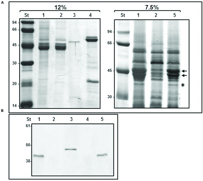
FIGURE 5.
Purified HBBs of polar and lateral flagella. (A) 12 and 7.5% SDS-PAGE of purified flagella HBB. The black arrows show FlaA and FlaB polar flagellins. The asterisk shows a band which molecular weight correlate to MotY and MotX proteins. (B) Western blot analysis using A. hydrophila AH-3 FlgT antiserum (1:1,000). Size standard (St); polar flagella HBB of AH-3 (1), AH-3ΔflgT mutant (2); purified His6-FlgT protein (3); lateral flagella HBB of AH-3::flhA (4); and AH-3ΔflgT mutant complemented with pBAD33-FLGT grown under inducer condition (5).

