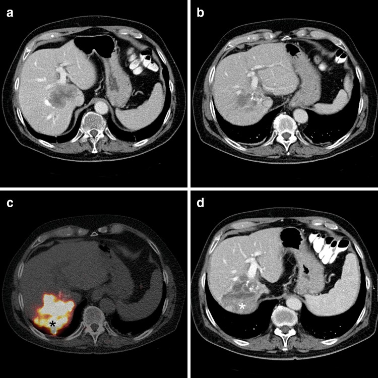
Fig. 1.
Induction of hypertrophy after 2 RE-treatments in a patient with CRLM. a CT scan prior to the first treatment with a CRLM located centrally in the right hemiliver, also involving the caudate lobe. b Three months after a whole liver treatment a decrease in the lesion size is seen. Segment 2–3 have hypertrophied (degree of hypertrophy: 16 %). c 90Y-PET/CT after a second selective treatment with glass microspheres (8 months after the first RE treatment): an intense accumulation of 90Y is seen in the lesion (*). d CT scan 2 months after the second treatment. The lesion in the right hemiliver has further decreased in size. A wedge-shaped hypodense area surrounds the lesion, consistent with radiation changes of the surrounding parenchyma (corresponding to the normal parenchyma with intense 90Y uptake on c (*). The hypertrophy of segment 2–3 has increased (degree of hypertrophy: 25 %). Also, segment 4 has hypertrophied (degree of hypertrophy: 20 %)

