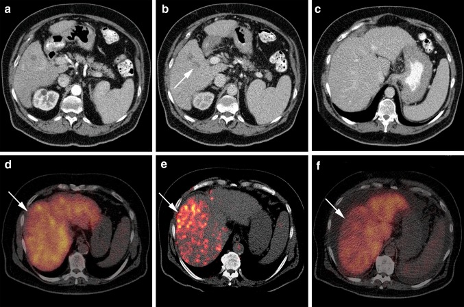
Fig. 2.
Decrease in 99mTc-mebrofenin uptake after right lobar 90Y-RE treatment. a A solitary, hypervascular lesion is present in segment 5 with wash-out (arrow) on the later obtained portal venous phase (b), consistent with an HCC. c The liver has a cirrhotic appearance (note the nodular surface). No lesions are seen elsewhere in the liver. d Hepatobiliary scintigraphy before RE-treatment shows a fairly homogeneous uptake of 99mTc-mebrofenin (cMUR: 3.0 %/min). e 90Y-PET/CT one day after right lobar treatment. 90Y has heterogeneously distributed in the right lobe with a higher dose in segment 4 and 8 (arrow in d, e and f). f Hepatobiliary scintigraphy 3 months after treatment. The uptake of 99mTc-mebrofenin is decreased in segment 4 and 8, corresponding to the area of higher 90Y deposit on the 90Y-PET/CT

