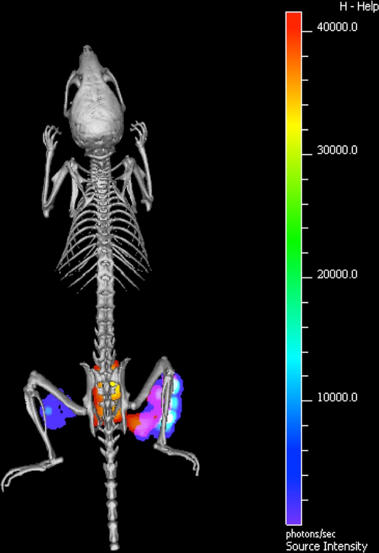
Fig. 1.
In vivo optical imaging of Staphylococcus aureus infection. Micro-computed tomography image of a mouse that was infected with a bioluminescent S. aureus strain in the right hind limb, and with a bioluminescent E. coli strain in the left hind limb. The NIR tracer vanco-800CW was administered intravenously [37]. The bioluminescence signal emitted by the infecting S. aureus and E. coli cells is depicted in rainbow scale, and the fluorescence signal due to vanco-800CW-binding in red–yellow scale. A clear co-registration of bioluminescence and NIR fluorescence was detected at the site of S. aureus infection. Moreover, a NIR fluorescence signal was detected in the bladder (in this image visible behind the spine). This bladder signal reflects the renal excretion of the tracer.
Reprinted with permission from [37]

