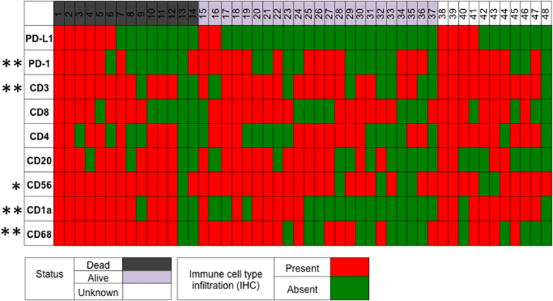
Figure 2. Expression of the immune checkpoint, PD-L1, is associated with the presence of tumor infiltrating lymphocytes and antigen presenting cells.
Each individual column represents a unique patient; of the 28 patients included in the study, 37 patients had known clinical outcomes, and 29 patients had both clinical outcome data and as well as demographic data available. Rows represent either PD-L1 expression or tumor infiltration by an immune cell type, as determined by immunohistochemistry. PD-L1 expression was significantly associated with infiltration by PD-1+ immune cells, CD3+ T cells, CD56+ natural killer cells, CD68+ cells, and CD1a+ dendritic cells. Presence of immune cells or positive PD-L1 expression was defined as IHC staining of >1% the tumor volume. * p < 0.05, **p < 0.01.

