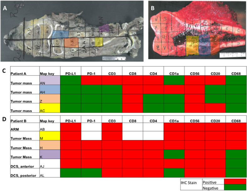
Figure 3. PD-L1 expression is consistent throughout the tumor mass.
(A,C) All four slides from the tumor mass of patient A did not demonstrate PD-L1 staining. Few immune cells are present in this tumor map. (B,D) All of the slides from the tumor mass of patient B stained PD-L1 positive with the exception of one section outside the tumor mass (not shown). Numerous immune cells are present throughout all sections of this tumor map. ARM = anterior resection margin, DCS = distal cross section.

