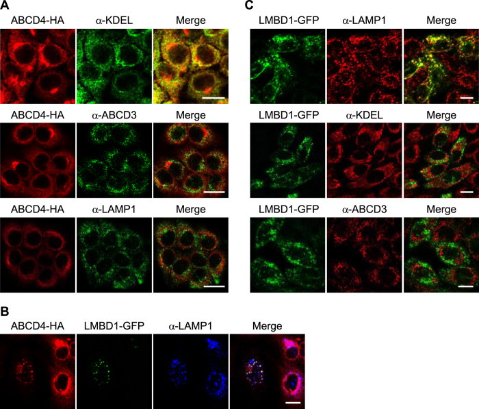
Figure 1. Subcellular localization of the ABCD4-HA and LMBD1-GFP expressed in mammalian cells.
(A) HuH7 cells stably expressing ABCD4-HA were stained with an anti-HA antibody. The ER, peroxisomes and lysosomes were labeled with an anti-KDEL, anti-ABCD3 and anti-LAMP1 antibody, respectively. Bar, 20 μm. (B) GFP-fused LMBD1 was transiently expressed in HuH7 cells stably expressing ABCD4-HA. The subcellular localization of ABCD4-HA and LMBD1-GFP was compared with that of lysosomes labeled with anti-LAMP1. Bar, 20 μm. (C) LMBD1-GFP was stably expressed in CHO cells. The distribution of LMBD1-GFP was compared with that of lysosomes, ER and peroxisomes stained with anti-LAMP1, anti-KDEL or anti-ABCD3, respectively. Bar, 10 μm.

