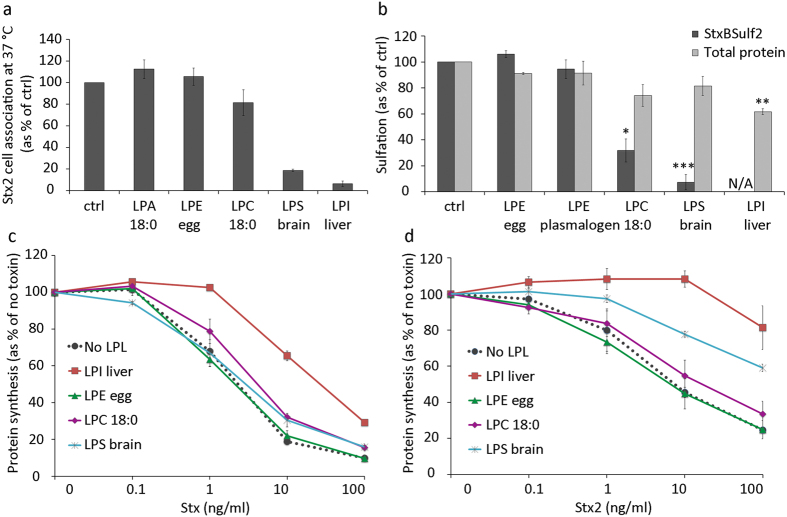
Figure 4. LPL effects on toxin cell association, transport and toxicity.
(a) HEp-2 cells were pretreated with 10 μM LPLs with acyl chains consisting of pure or mostly C18:0 for 30 min, prior to incubation with 125I-Stx2 for 20 min at 37 °C. Cells were washed, lysed and the radioactivity was measured. Cell-associated 125I-Stx2 is expressed as % of control (mean ± deviation; n = 2). (b) Sulfation assay to measure the amounts of StxBSulf2 transported to the Golgi apparatus where the modified toxin is sulfated. HEp-2 cells were pre-incubated with 35SO42− for 1.5 h and then incubated with 10 μM LPLs with acyl chains consisting of pure or mostly C18:0 for 30 min, followed by addition of 2 μg/ml StxBS2 for 1.5 h. Shiga toxin was immunoprecipitated from cell lysates and radioactivity was counted. Results are shown as % of ctrl (mean ± SEM; n = 3). N/A stands for not applicable (below detection limit). (c,d) Toxicity assay with Shiga holotoxin (Stx) and Stx2. Cells were pretreated with 5 μM LPLs with acyl chains consisting of pure or mostly C18:0 for 30 min, followed by incubation with tenfold serial toxin dilutions for 4 h in leucine-free medium. Protein synthesis was measured and results are shown as % of no toxin (mean ± deviation; n = 2 for each of the toxins).

