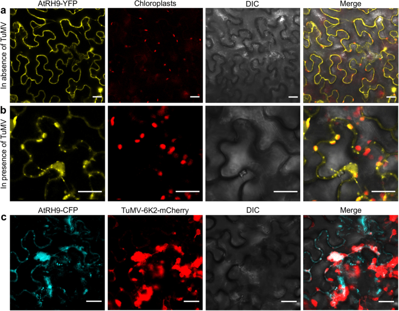
Figure 3. Altered Localization of AtRH9 in Arabidopsis during TuMV Infection.
(a) Transient expression of AtRH9-YFP in N. benthamiana leaf epidermal cells. YFP fluorescence was observed using a confocal microscope 48 hours post agroinfiltration (hpa). (b) AtRH9-YFP was expressed in N. benthamiana leaves infected by TuMV::6K2-mCherry. Localization of AtRH9 is associated with chloroplasts in the course of viral infection at 72 hpa. Bars, 20 μm. (c) Transient expression of AtRH9-CFP in N. benthamiana leaves infected by TuMV::6K2-mCherry. CFP fluorescence was observed using a confocal microscope 72 hpa. AtRH9-CFP was observed mainly with irregularly-shaped globular-like aggregations from red fluorescence from TuMV VRCs (6K2-mCherry). DIC, differential interference contrast. Bars, 20 μm.

