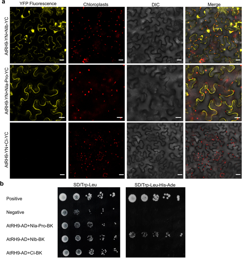
Figure 4. Protein-Protein Interactions between AtRH9 and TuMV Viral Proteins.
(a) BiFC assay for interactions between AtRH9 and TuMV NIb, NIa-Pro. N. benthamiana leaves were co-agroinfiltrated with constructs expressing AtRH9 with TuMV NIb, NIa-Pro and CI fused to the N- and C- terminal half of YFP, respectively. The reconstructed YFP fluorescence was recorded 48 hours post agroinfiltration (hpa) by confocal microscopy. DIC, differential interference contrast. Bars, 20 μm. (b) Yeast two-hybrid assay for protein-protein interaction between AtRH9 and NIb. Growth of yeast cells co-transformed with pGAD-AtRH9 and pGBK-NIb, pGBK-NIa-Pro or pGBK-CI were placed on synthetic medium lacking Tryptophan and Leucine (SD/Trp-Leu) to confirm the correct transformation. Different dilutions of yeast transformants were spotted onto a high-stringency selective medium (SD/Trp-Leu-His-Ade) to detect positive interactions, respectively. Co-transformation of pGAD-VPg with pGBK-eIF(iso)4E or pGAD-AtRH9 with pGBK empty vector were used as the positive or negative controls, respectively.

