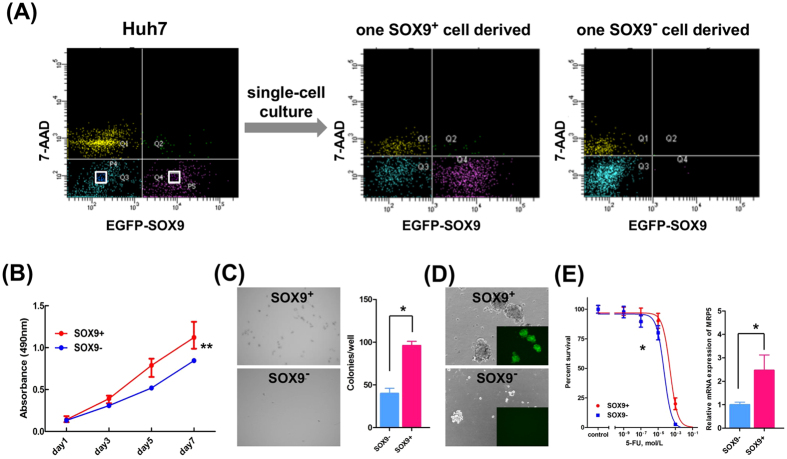
Figure 1. Cancer stem cell properties of SOX9+ Huh7 cells in vitro.
(A) Single-cell culture of SOX9+ and SOX9− Huh7 cells. FACS analyses revealed that isolated SOX9-EGFP+ cell differentiated both EGFP+ and EGFP− cell fractions (middle panel), whereas isolated SOX9-EGFP− cell only to EGFP− cell fraction (right panel). (B) Cell proliferation assays showed SOX9+ cells proliferate more than SOX9− cells (repeated-measures ANOVA, **P < 0.01). (C) Microscopic appearance and the colony numbers in the anchorage-independent growth assay (Student’s t-test, *P < 0.05). (D) Phase-contrast images in the sphere-forming assay. (E) IC50 of 5-FU in SOX9+ and SOX9− cells (left panel, F-test, *P < 0.05) and qRT-PCR analyses of MRP5 in SOX9+ and SOX9− cells (right panel, Student’s t-test, *P < 0.05).

