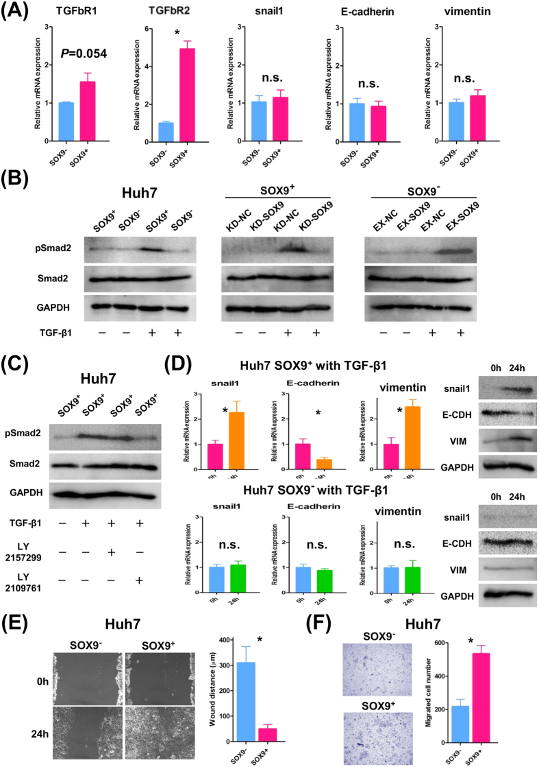
Figure 3. TGFb-induced EMT phenotype in SOX9+ Huh7 cells.
(A) qRT-PCR analysis of TGFb receptors and EMT-related genes in SOX9+ and SOX9− cells without TGFb stimulation (Student’s t-test, *P < 0.05). Data are shown as the mean ± SD. (B) Western blot analysis showed TGFb-induced pSmad2 upregulation in SOX9+ but not in SOX9− cells (left panel), and regulation of TGFb-induced pSmad2 upregulation by SOX9 (middle and right panel). (C) Western blot analysis showed TGFb-induced pSmad2 upregulation in SOX9+ cells was suppressed by LY2109761 but not LY2157299. (D) qRT-PCR and western blot analyses showed elevation of snail1 and vimentin accompanied by E-cadherin downregulation, caused by TGFb stimulation in SOX9+ cells (Student’s t-test, SOX9+ Huh7 cells; *P < 0.05, SOX9− Huh7 cells; not significant). Data are shown as the mean ± SD. (E) Wound-healing assay of SOX9+ and SOX9− cells with TGFb stimulation (Student’s t-test, *P < 0.05). (F) Migration assay of SOX9+ and SOX9− cells with TGFb stimulation (Student’s t-test, *P < 0.05).

