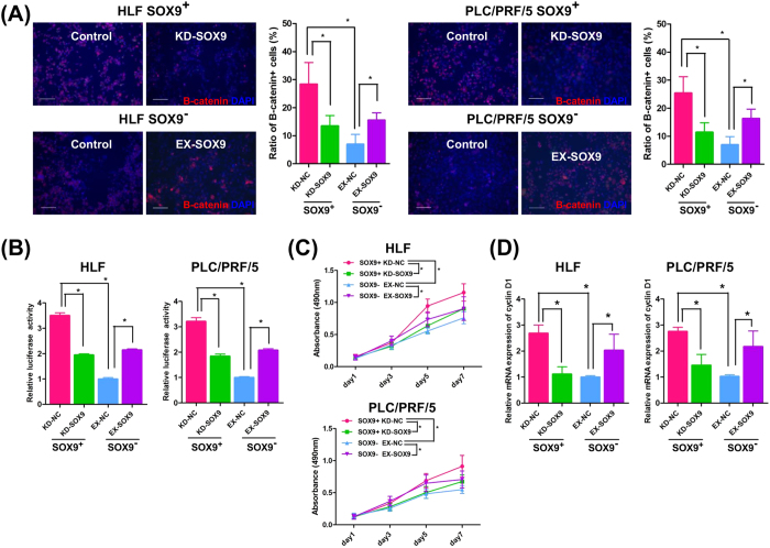
Figure 4. SOX9 activates the Wnt/beta-catenin pathway in HLF and PLC/PRF/5 cells.
(A) Beta-catenin staining. Note the nuclear localized, activated beta-catenin expression in the steady state SOX9+ cells (Control) and its reduction by SOX9 knockdown (KD-SOX9). Exogenously introduced SOX9 (EX-SOX9) accelerated beta-catenin activation in SOX9− cells (Student’s t-test, *P < 0.05). Scale bar represents 100 μm. (B) TCF/LEF luciferase assay with SOX9-gain/loss of function experiments (Student’s t-test, *P < 0.05). (C) Cell proliferation assays with SOX9-gain/loss of function experiments (repeated-measures ANOVA, *P < 0.05). (D) qRT-PCR analysis of cyclin D1 with SOX9-gain/loss of function experiments (Student’s t-test, *P < 0.05). Data are shown as the mean ± SD. KD-NC; control knockdown, KD-SOX9; SOX9 knockdown, EX-NC; control overexpression, EX-SOX9; SOX9 overexpression.

