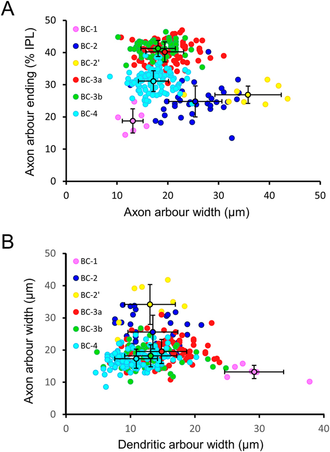
Figure 1. Morphological differentiation of OFF BC groups.

(A) The distribution of BC axonal arbour ending depth in the IPL versus its horizontal extension displays a separation of all OFF BC groups, except for BC-3a and 3b. Notably, the classification of BC-2′ as a separate group based on electrophysiological criteria, is supported by its wider axonal arbour compared to BC-2. (B) Plotting axon arbour versus dendritic arbour width clearly distinguishes BC-1 from BC-2 and the other BC cell types. Black circles and bars indicate the average ± s.d.
