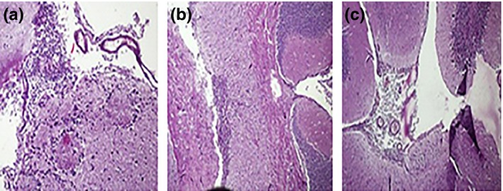
Figure 3.

Representative histopathological micrographs of mice brain sections after 12 weeks of infection: (a) deeper brain tissue inflammatory reaction, (b) oedema of brain tissue beneath choroid plexus and (c) cerebellum part of brain with lymphocytic granuloma.
