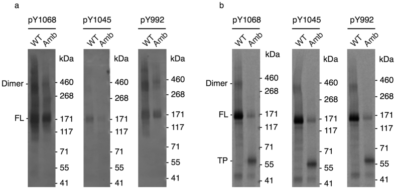
Figure 3. In vitro phosphorylation of selected tyrosine residues.
(a) Immunoblotting of EGFR-eYFP (WT) and mutant with incorporated AzF (Amb) against phosphotyrosine 1068 (pY1068), 1045 (pY1045) and 992 (pY992) using specific antibodies. (b) Autoradiography of corresponding blotting membranes. Dimers as well as the full-length protein (FL) and the termination product (TP) are indicated. Blots (a) and autoradiograms (b) have been adapted in contrast, brightness and sharpness for better visibility. The original images can be found in supplementary Fig. 2.

