
Figure 5. OCT imaging of corneal morphology in later post-mortem intervals.
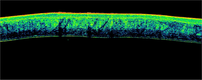
A horizontal, high-resolution Fourier-domain, cross-sectional OCT scan of the central cornea (2 mm of diameter). Irregular variations in corneal contour due to corneal edema, ocular hypotony or advanced decomposing processes were observed during OCT scans performed after 48 hours from death. These findings probably led to an increase in variability of pachymetric measurements obtained during later post-mortem intervals (within a range of 48–96 hours) compared with earlier results.