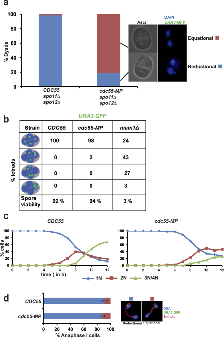
Figure 4. cdc55-MP affects reductional segregation of chromosomes during meiosis I in spo11Δ spo12Δ cells but not in wild type cells.
(a) CDC55 spo11Δ spo12Δ and cdc55-MP spo11Δ spo12Δ cells containing heterozygous GFP-tagged URA3 sequences were induced to enter meiosis by transferring them into SPM followed by incubation at 30 °C for 24 h. Cells were fixed and stained with DAPI to visualize DNA and segregation of GFP-tagged URA3 dots was assayed by fluorescence microscopy (N = 200). Representative images of dyads showing equational and reductional segregation of URA3-GFP sequences are depicted on the right. (b) Detection of homozygous URA3-GFP and DNA in tetrads produced by CDC55, cdc55-MP and mam1Δ strains. Tetrads produced by the strains were dissected onto YPD plates and grown at 30 °C. Spore viability (n = 100) was scored after 3 days. (c,d) Analysis of meiosis in CDC55 and cdc55-MP cells containing heterozygous URA3-GFP. Samples of cultures at indicated time points were taken out, fixed and stained with DAPI and anti-tubulin antibodies. (c) Kinetics of meiotic nuclear division shown by percentage of mononucleate, binucleate and tri/tetranucleate cells during the time course. (d) Proportion of cells containing anaphase I spindles undergoing reductional and equational segregation of URA3-GFP dots are depicted (N = 100 in triplicates). Difference in the frequencies of reductional segregation between CDC55 and cdc55-MP strains is not statistically significant (Student’s t-test, P < 0.1). Data shown are representative of 3 independent experiments.

