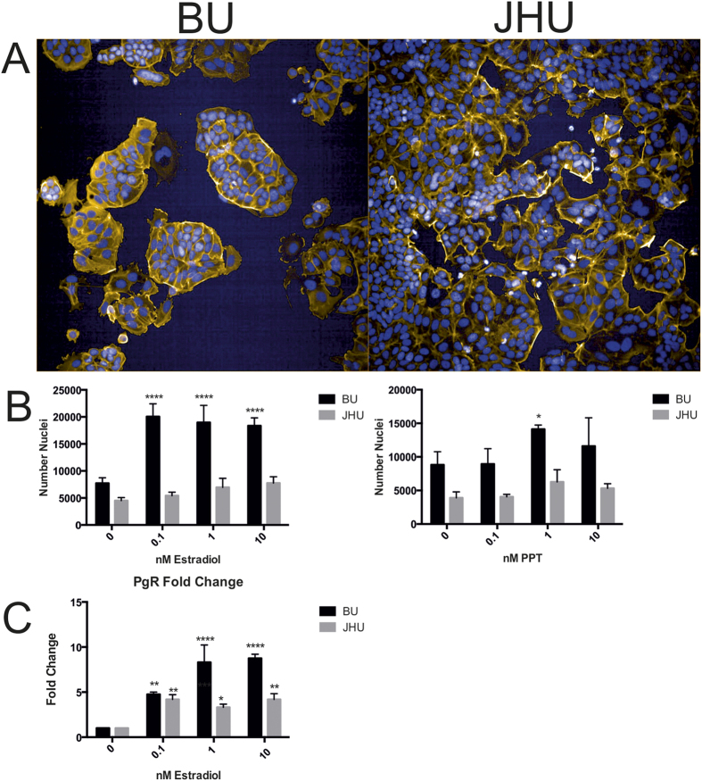
Figure 1. Morphological, phenotypical and gene expression differences of MCF-7 cells from the same ATCC batch (passsage number 154 JHU & passage number 150 BU).
(A) MCF-7 sublines (JHU & BU) display distinct morphological differences. At 0 hours, BU MCF-7 cells and JHU cells have unique morphologies. MCF-7 cells grown and expanded at BU grow in large aggregations while JHU cells grown in the BU laboratory are flat, with cobblestone morphology. (B) Following 72 hours of exposure to estradiol (E2), BU MCF-7 cells displayed significant increases in proliferation (cell count) at concentrations of 0.1, 1 and 10 nM, while JHU cells did not have a significant change in cell count (left). Exposure to the estrogen receptor alpha agonist propyl pyrazole triol (PPT) for 72 hours resulted in a significant increase in proliferation at concentrations of 0.1, 1.0 and 10 nM in the BU subline, while JHU cells did not exhibit significant changes (right). (C) Gene expression analysis of the estrogen receptor target progesterone receptor (PgR) following 6 hours of exposure to E2 indicated that BU MCF-7 cells are responsive to low levels of estrogen. All experiments have been performed in one laboratory (BU) by one individual to exclude any possible inter-laboratory and/or inter-operator effects on reproducibility. *p < 0.05, **p < 0.01, ***p < 0.001, ****p < 0.0001.

