Fig. 2.

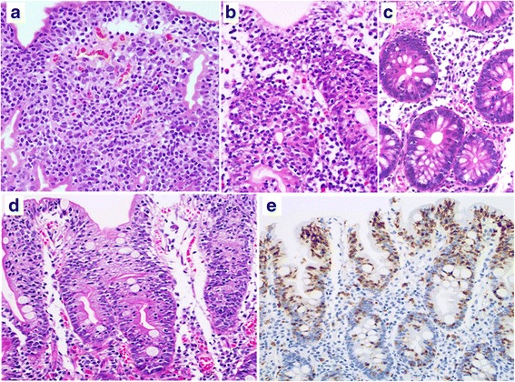
Patient 1 (a–c). a: A diffuse infiltrate of small to medium-sized atypical lymphocytes and many atypical IELs are seen in the duodenal (a) and cecal (b) mucosa. c: Small intraepithelial lymphocytes are scattered in the non-neoplastic ascending colonic mucosa, indicating lymphocytic colitis. Patient 2 (d, e). d: Duodenal enteropathy with atrophic villi and increased IELs. e: Many small CD8-positive IELs are seen in the duodenum (a, b, c, d: H&E stain; e: immunohistochemistry, hematoxylin stain)
