
Fig. 4.
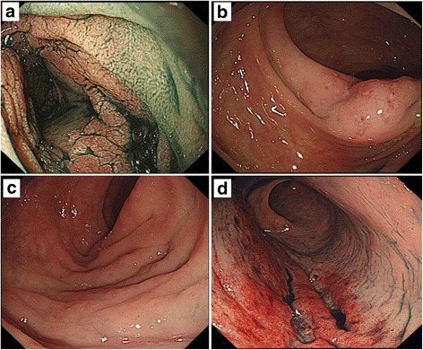
Patient 3 (a–d). a: Upper GI endoscopic view of the duodenal second portion shows an edematous mucosa with a nodular or mosaic pattern. b: A lower GI endoscopic view of the ascending colon shows an edematous mucosa with multiple erosions. Lower GI endoscopic views show an edematous mucosa in the sigmoid colon (c) and reddish longitudinal ulcers in the rectum (d)