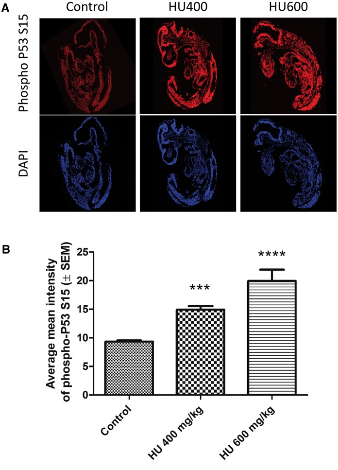
FIG. 4.
Hydroxyurea exposure induced a widespread increase in phospho-P53 immunoreactivity. A, Representative multiphoton and confocal microscopy images taken at × 20 of whole embryo sections showing increasing phospho-P53 immunoreactivity (red, top panel) and the DAPI nuclear counterstain (blue, bottom panel). B, Average mean intensity of phospho-P53 immunoreactivity in whole embryo sections. A significant increase in phospho-P53 intensity was detected in the HU400 and HU600 groups. N = 4–5 for each treatment group. ***P < .001, ****P < .0001, one-way ANOVA with Bonferroni post-hoc test.

