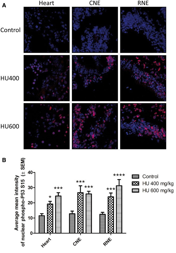
FIG. 5.
Nuclear translocation of phospho-P53 increased significantly with hydroxyurea treatment. A, Representative multiphoton and confocal images of phospho-P53 immunoreactivity taken at × 63 magnification. Three tissues were analyzed per embryo across all treatment groups (Heart, CNE: caudal neuroepithelium, RNE: rostral neuroepithelium). B, Quantification of phospho-P53 colocalization with DAPI. N = 4–5 for each tissue and treatment group. *P < .05, ***P < .001, ****P < .0001 compared with control, 2-way ANOVA with Bonferroni post-hoc test. There were no statistically significant differences in phospho-P53 nuclear translocation between tissues in the embryo, and no statistically significant interaction between the treatment and tissue variables.

