Figure 1.
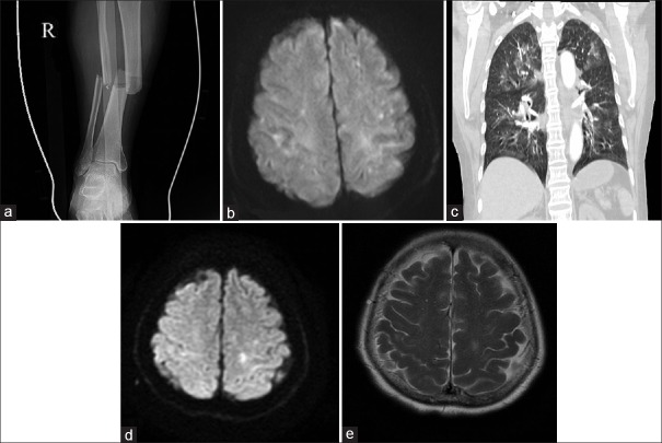
(a) Radiograph of the right leg showing tibia and fibula fractures. (b) Four hours after the accident, diffusion-weighted magnetic resonance imaging of the brain showed some punctate foci of restricted diffusion in a starfield pattern. (c) Twenty hours after the accident, computed tomography angiography of the chest showed bilateral ground-glass opacities, without pulmonary embolism. Ten days after the accident, diffusion-weighed magnetic resonance imaging (d) and T2-weighted magnetic resonance imaging (e) of the brain showed a high-signal focus in the left semioval center.

